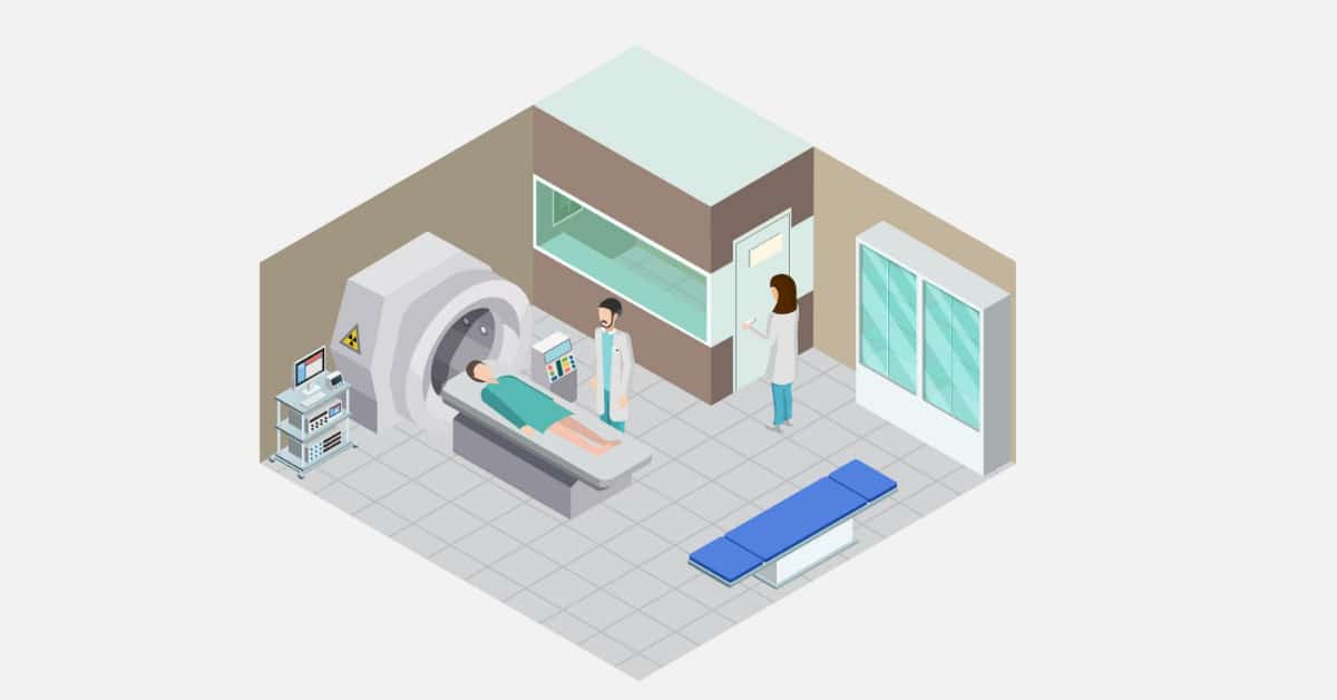
The discovery of X-rays is one of the great discoveries in medicine that changed a lot in the quality of diagnosis and treatment that the patient receives. X-rays enabled doctors and scientists to see inside the human body and determine the cause and source of pain.
With the development of medicine, so did the field of medical imaging. Today, thanks to nuclear medicine, we do not see only pictures of the human body’s organs, but we are beginning to see how these organs perform their vital functions inside the human body.
We are now seeing what happens to the molecules and cells that perform their functions inside the human body in vivo without hindering them from their function.
One of the benefits of this field is the speed of diagnosis of the disease and sometimes even before symptoms of the disease occur, and this is when pathological changes occur at the level of cells and molecules before the organ is completely affected.
Nuclear Medicine Uses Some Radioactive Materials (Radioisotopes)
Nuclear medicine uses some radioactive materials (Radioisotopes) to diagnose and treat many diseases. It is considered a painless and very safe examination for the patient, which is performed with a small amount of radiation, so there is no need for any concern or fear of the examination.
The reason for naming nuclear is due to the fact that the rays used come from the nucleus of the atom. The idea of nuclear medicine in general is to give the patient a radioactive substance that is concentrated in one of the organs of the human body, and then the patient is photographed by a special camera that captures the radiation coming out of the organ, and so we can know what happens inside the patient’s body.
The amount of dose of the radioactive material depends on the type of examination and the age of the patient. The most common tests in nuclear medicine are bone, kidney, thyroid, heart, lungs, brain, lymphatic system, etc.
Why Nuclear Medicine?
Through nuclear medicine, we can obtain information and details about diseases and the human body that we may not be able to obtain in another way or with another device.
Nuclear medicine is distinguished from other types of radiography in that it can visualize the function of the body’s organs, not just anatomy.
Other types of radiography mostly focus only on anatomy. For example, kidney, CT scans and sound waves can visualize the anatomy of the kidney.
With them, we can see the shape of the kidney and measure its dimensions and whether there is a change in its shape, which indicates the presence of illnesses from diseases and others. In nuclear medicine, we can see the function of the kidney, its efficiency, and so on.
Another difference between nuclear medicine and the other types is the radiation source. The patient in medical imaging devices receives radiation from an x-ray machine, so the radiation source is external. Whereas in nuclear medicine the patient himself is the source of radiation after he is given a radioactive substance.
Imaging Method in Nuclear Medicine
In usual diagnostic radiology, the energy source is outside the patient, such as an x-ray tube, a magnetic field, or even sound waves, and this energy is represented by a picture that shows the the bones and tissues of the patient.
In nuclear medicine, the energy source is the patient himself, by injecting, drinking, or inhaling a radioactive material, and then this radioactive material is photographed by a gamma camera, SPECT or PET device. But how is this radioactive material sent to the place to be photographed? This is done by pharmaceutical ability to go to the organ to be imaged. For example, MDP, when injected into the human body, is deposited in the bones.
If we want the radioactive material to also be concentrated in the bones, then we mix it with MDP and it goes and the radioactive material is concentrated in the bones. The pharmaceutical substance mixed with a radioactive substance is called Radio-pharmaceutical. So the pharmaceutical substance is only a carrier of the radioactive material to the organ to be photographed.
How to Give the Radioactive Material to the Patient?
Mostly the patient is given the radioactive material by injection into the vein, but there are other ways to take the radioactive material, for example by mouth (capsule or syrup) or even by inhaling the radioactive substance that is in the form of gas.
Types of Radioactive Materials
There are a group of radioisotopes, each of which has different half-lives, and different radiological and chemical properties that can be used to detect specific diseases in the body.
1.Technetium-99M: one of the most commonly used radioisotopes in diagnosis, accounting for more than 80% of the tests. Bone scan with radioactive technetium.
2.Iodine-123: Diagnosis, especially thyroid gland, and neurological diseases (such as Alzheimer’s and Parkinson’s disease).
3.Iodine-131: Treatment of thyroid diseases, including cancer. Radioactive iodine treatment
4.Fluorine-18: Imaging of malignancies.
5.Thallium-201: Heart monitoring during exercise.
6.Gallium-67: Imaging of tumors and regeneration of inflammation.
7.Indium-111: Identification of blood clots, infections and rare cancers.
8.Xenon 133: Is aspirated to study lung ventilation.
9.Chromium 51: Follow-up of blood cells, diagnosis of gastrointestinal bleeding.
10.Lutetium-177: Diagnosis and treatment of small cancer tumors.
11.Yttrium-90: Treating cancer and relieving pain in arthritis.
Possible Risks and Side Effects
The amount of radiation in nuclear medicine examinations is very small and is not a cause for concern. This does not mean that there are no risks of exposure to radiation, but it is few like the rest of the other radiological examinations. More about the risks of radiation in this article click here.
Also, it is good in nuclear medicine examinations that the risks of allergy to the human body (sensitivity) from the radioactive pharmaceutical substance are very small, and even rare compared to the dye used in CT and MRI scans. So crosses are very safe.
Contraindications to the Examination
Nuclear medicine scans are performed for all age groups, from children and newborns to the elderly. With the exception of pregnant women, the examination is postponed until after childbirth, because radiation may cause harm to the fetus. And when there is suspicion of a pregnancy, a pregnancy test is performed to ensure that it is not present.
Preparing for the Examination
Usually the examination does not require preparation, but the patient may sometimes be asked to be fasting, and he may be asked to stop using some medicines or the use of certain medicines, so the instructions given when taking the appointment must be adhered to.
Post-examination Instructions
The patient must drink sufficient quantities of fluids and go to the toilet continuously to speed up the process of the body getting rid of the radioactive material through urine. A nursing mother must stop breastfeeding for a certain period due to the presence of radiation with breast milk, and she will be informed of the period during which she must stop breastfeeding by the staff of the nuclear medicine unit. After the examination, the patient can go about his daily life normally.


